10 May 2017
By Bryan T. Smyth
bryan@TheCork.ie
Tyndall Photonics Researchers Working with EU Partners to Develop Prototype
Researchers at Tyndall National Institute, Cork, are taking part in a new European project to develop a low cost, easy to use 3D imaging system that will help enable faster and more accurate diagnosis of colorectal cancers. The Optical Coherence Tomography (OCT) system is being developed by Tyndall as part of the recently launched PICCOLO project, an EU Horizon 2020 funded initiative, bringing together scientists and clinicians across Europe to create an endoscope probe to better diagnose bowel polyps and early colon cancer.

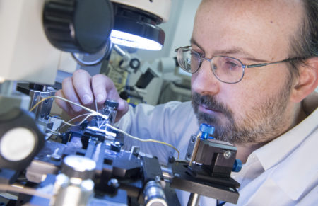
Laser researcher Dr Brendan Roycroft, aligning a new multi-contact tunable laser in Tyndall National Institute. The laser can provide information from different depths in human tissue without damaging it in any way, creating a high resolution 3D image. Photograph by Gerard McCarthy.
Tyndall researcher, Dr. Brendan Roycroft explains: “Using breakthrough photonics, (the generation, manipulation and utilisation of light), we can create 3D images to diagnose non-cancerous polyps, pre-cancerous polyps and early colon cancers via an endoscope. The imaging is non-destructive, and we will be able to see below the surface of the tissue to identify internal polyp structure, providing valuable information as to the polyp type without a surgeon having to cut out a non-cancerous polyp and examine it in a lab. This will result in less discomfort for the patient, while also reducing the costs and time necessary for accurate diagnosis.”
Colorectal cancer represents around one tenth of all cancers worldwide. Through early, accurate diagnosis and precise intervention, cure rates can increase by up to 90%. Across the globe, colon cancer remains the second most common cancer in women, behind breast cancer, and the third most common in men, behind lung and prostate cancer.
Tyndall’s OCT research team includes researchers from Cork Institute of Technology and the Irish Photonic Integration Centre (IPIC) hosted at Tyndall. The advanced PICCOLO endoscope will use OCT, MPT (Multi-Photon Tomography) and fluorescence imaging to provide high-resolution information, giving details of the changes occurring at the cellular level. The PICCOLO project team aim to have refined their first prototype by the end of 2018 and target clinical trials to begin around 2020. It is hoped the new image based diagnosis method could eventually be applied to diseases in other organs of the body.


